



Complete tooth loss reshapes far more than just a person’s smile; it fundamentally alters facial structure, digestion, and self-confidence. Historically, removable acrylic dentures served as the only viable recourse for severe edentulism. Yet, these traditional appliances often bring their own set of daily frustrations, ranging from embarrassing slippage during meals to localized tissue irritation and ongoing bone loss. The restorative dentistry landscape has evolved dramatically over the last two decades, shifting its focus toward highly secure, permanent alternatives that closely mimic natural human physiology.
At the forefront of this shift is a strategic surgical approach that maximizes existing bone volume to securely support a complete arch of teeth. Specialized institutions like Dentprime frequently utilize these advanced protocols to deliver highly stable, long-lasting outcomes for patients looking to completely bypass the inconveniences of traditional prosthetics.
The Architectural Ingenuity of Strategic Placement
Unlike replacing individual teeth with single, isolated titanium posts, restoring a full upper or lower jaw does not require an implant for every missing tooth. The methodology behind all-on-4 dental implants relies heavily on precise mathematical load distribution and structural engineering. Dental surgeons embed exactly four titanium fixtures into the jawbone, which then serve as a robust, immovable foundation for a custom-crafted bridge.
The true ingenuity of this treatment lies in the specific positioning and trajectory of these fixtures. While the two anterior (front) posts are inserted vertically into the jaw, the two posterior (back) posts are tilted at a distinct angle typically between 30 and 45 degrees. This angular placement dramatically increases the bone-to-implant contact area, allowing a relatively small number of anchors to support the immense occlusal forces generated during chewing.
Bypassing Anatomical Roadblocks
This tilted posterior placement is not merely a structural choice; it effectively solves one of the most common medical hurdles in restorative surgery: severe jawbone atrophy. When teeth are missing for extended periods, the jawbone naturally shrinks due to a lack of stimulation. In the upper jaw, this resorption causes the sinus cavities to expand downward. In the lower jaw, the shrinking bone brings the alveolar ridge dangerously close to the mandibular nerve.
By tilting the back implants, surgical specialists can utilize the dense, high-quality bone located further toward the front of the mouth. This clever trajectory completely bypasses the sinus cavities and sensitive nerve canals. Consequently, many individuals who were previously told they lacked the bone density for implants or who faced the prospect of invasive, months-long bone grafting procedures can now receive permanent restorative care without those preliminary surgeries.
Precision Through Digital Mapping
Executing this intricate procedure requires absolute precision, moving far beyond traditional two-dimensional x-rays. Modern practitioners rely heavily on advanced digital imaging, specifically Cone Beam Computed Tomography (CBCT). This technology captures highly detailed, three-dimensional cross-sections of the patient’s craniofacial anatomy.
Using this data, dentists map out the exact trajectory, depth, and placement of each titanium screw within specialized software long before the actual surgery takes place. This digital workflow often culminates in the creation of a 3D-printed surgical guide, which sits over the patient’s gums during the procedure to ensure the implants are placed exactly where they were digitally planned, minimizing surgical trauma and accelerating recovery times.
The Concept of Immediate Functionality
The procedural timeline represents another significant leap forward for patient experience. Traditional implantology often required patients to wait anywhere from three to six months for the titanium to fuse with the bone, a biological process known as osseointegration before attaching any functional teeth.
The multi-anchor system inherently provides extreme primary stability, frequently allowing for immediate loading. On the very same day of the surgical insertion, a temporary, fully functional set of teeth is firmly attached to the newly placed implants. Patients enter the facility with failing dentition or unstable dentures and leave hours later with a fixed, aesthetically pleasing smile. This provisional bridge securely manages necessary chewing forces while the underlying bone heals and solidifies its grip on the titanium over the subsequent months.
Crafting the Permanent Prosthetic
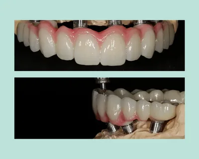
Once the biological healing phase reaches completion, the temporary bridge is swapped for the final, custom-manufactured prosthetic arch. The materials chosen for this permanent fixture dictate both its physical durability and its visual authenticity. Advanced dental laboratories typically mill these final restorations from ultra-strong, biocompatible materials such as monolithic zirconia, or they utilize a lightweight titanium framework layered with high-grade dental porcelain.
These materials resist micro-fractures and completely repel stains, all while beautifully replicating the translucent, light-scattering properties of organic human enamel. Furthermore, the prosthetic flawlessly replaces the lost gingival tissue, utilizing customized pink-hued ceramics to ensure a seamless, undetectable transition at the natural smile line.
Maintenance and Long-Term Viability
Securing a permanent prosthetic demands a dedicated, lifelong hygiene routine to protect the underlying foundation. While the synthetic teeth and titanium posts cannot develop cavities, the biological soft tissues surrounding the implants remain highly vulnerable to bacterial buildup. If plaque is allowed to accumulate beneath the bridge, it can trigger peri-implantitis, an aggressive inflammatory condition capable of eroding the supporting jawbone. Standard toothbrushing alone cannot adequately clean these complex structures. Patients must integrate specific hygiene tools, such as high-pressure water irrigators and specialized interdental floss threaders, to flush out microscopic debris trapped between the prosthetic and the gum tissue. Consistent professional cleanings and routine radiological examinations are paramount to verifying the structural integrity of the hardware, ensuring the restoration performs optimally for decades.